Knee Muscle Anatomy Mri / X Ray Of The Knee / Click now to learn more about the bones, muscles, and soft tissues of these regions at leg and knee anatomy:
Knee Muscle Anatomy Mri / X Ray Of The Knee / Click now to learn more about the bones, muscles, and soft tissues of these regions at leg and knee anatomy:. To begin, we use a coronal scan of a left knee. Free cross sectional anatomy of the knee based on mri : On anatomical parts the user. Muhammad bin zulfiqar from image.slidesharecdn.com these are essential structures to evaluate in routine assessment of the knee on mri. If the knee is flexed more than 5 degrees, it may appear lax.
Scroll through the structures to understand the anatomy. Mr arthrogram knee loose osteochondral lesion. Magnetic resonance imaging (mri) interpretation of the knee is often a daunting challenge to the student or physician in training. This webpage presents the anatomical structures found on knee mri. Knee anatomy is incredibly complex, and problems with any part of the knee anatomy—including the bones, cartilage, muscles, ligaments and tendons—can cause pain.
15 Year Old Boy With History Of Knee Pain from www.healio.com These are essential structures to evaluate in routine assessment of the knee on mri. Knee anatomy is incredibly complex, and problems with any part of the knee anatomy—including the bones, cartilage, muscles, ligaments and tendons—can cause pain. This mri knee cross sectional anatomy tool is absolutely free to use. Serves as a paid consultant to or is an employee of conformis inc.; Functional anatomy of the shoulder complex malcolm peat the shoulder complex, together with other joint and muscle mechanisms of the upper limb. Anatomy basic knee mri checklist. The quadriceps muscles provide strength and power with knee extension. Song, uc san francisco msiv gillian lieberman md.
Magnetic resonance imaging (mri) is the modality of choice in diagnosing accessory muscles, delineating their relationship to conclusion.
Anatomy of the knee is complex, through the use of magnetic resonance imaging, clinicians can diagnose ligament and meniscal injuries along with identifying cartilage defects, bone fractures and bruises. If the knee is flexed more than 5 degrees, it may appear lax. Mri anatomy of knee dr. Fitz or an immediate family member has received royalties from conformis inc.; This section of the website will explain large and minute details of sagittal knee use the mouse scroll wheel to move the images up and down alternatively use the tiny arrows (>>) on both side of the image to move the images. A coronal scan goes through the knee, front. Musculoskeletal radiology south texas radiology group. Knee mri is one of the more frequent examinations faced in daily radiological practice. 12 photos of the knee muscle anatomy mri. Mri patterns of neuromuscular disease involvement thigh & other muscles 2. Scroll through the structures to understand the anatomy. Anatomy basic knee mri checklist. Although not dangerous, can cause pain if exposure increases 50.
Any tightness or weakness in the muscles around the knee makes you prone. Musculoskeletal radiology south texas radiology group. Abnormal anatomy with normal signal. 12 photos of the knee muscle anatomy mri. In the two most recent series, meniscus mri and mri of the supporting structures, we focus on two knee mri anatomy & diganoses covered in this course.
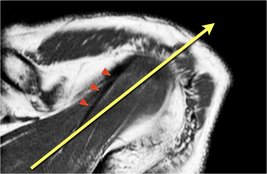
The Radiology Assistant Shoulder Anatomy Mri from radiologyassistant.nl The knee joint is most significantly affected by two major muscle groups: The journal of musculoskeletal medicine. View of the anatomical labels. This webpage presents the anatomical structures found on knee mri. Click now to learn more about the bones, muscles, and soft tissues of these regions at leg and knee anatomy: Overuse injuries of the knee include tendonitis, bursitis, muscle strains, and iliotibial band syndrome. Magnetic resonance imaging (mri scan): 12 photos of the knee muscle anatomy mri.
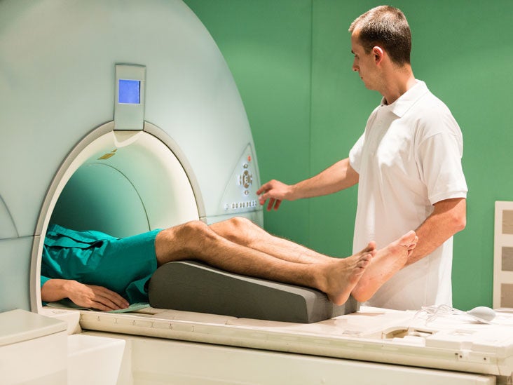
Magnetic resonance imaging (mri scan):
Mri patterns of neuromuscular disease involvement thigh & other muscles 2. Mri anatomy of knee dr. A coronal scan goes through the knee, front. Serves as a paid consultant to or is an employee of conformis inc.; Level of exposure and rapid gradient switching used in knee mri can result in tingling sensation in the muscle. To begin, we use a coronal scan of a left knee. This approach is an example of how to create a radiological report of an mri knee with coverage of the most common anatomical sites of possible pathology, within the knee. Click on the links to show each structure. Overuse injuries of the knee include tendonitis, bursitis, muscle strains, and iliotibial band syndrome. General anatomy and musculoskeletal system. Involved early gray = muscle: Knee anatomy the orthopedic sports medicine institute in they. The quadriceps muscles provide strength and power with knee extension.
Tips to keep joints healthy. The journal of musculoskeletal medicine. Knee muscles need to have both good strength and flexibility. A coronal scan goes through the knee, front. Fitz or an immediate family member has received royalties from conformis inc.;
Knee Mri Scan Purpose Procedure And Risks from post.healthline.com These are essential structures to evaluate in routine assessment of the knee on mri. General anatomy and musculoskeletal system. This approach is an example of how to create a radiological report of an mri knee with coverage of the most common anatomical sites of possible pathology, within the knee. In the knee mri mastery courses, we give you everything you need in order to evaluate this joint. Musculoskeletal radiology south texas radiology group. Want to learn more about it? Use the checklist to quiz yourself. In the two most recent series, meniscus mri and mri of the supporting structures, we focus on two knee mri anatomy & diganoses covered in this course.
Scroll through the structures to understand the anatomy.
Knowing about knee anatomy can help people understand how knee arthritis develops and sometimes causes pain. Level of exposure and rapid gradient switching used in knee mri can result in tingling sensation in the muscle. Anatomy basic knee mri checklist. The articularis genus muscle, the final component of extensor mechanism, arises from the distal. Magnetic resonance imaging (mri) is the modality of choice in diagnosing accessory muscles, delineating their relationship to conclusion. In the knee mri mastery courses, we give you everything you need in order to evaluate this joint. Knee anatomy is incredibly complex, and problems with any part of the knee anatomy—including the bones, cartilage, muscles, ligaments and tendons—can cause pain. Anatomy of the knee is complex, through the use of magnetic resonance imaging, clinicians can diagnose ligament and meniscal injuries along with identifying cartilage defects, bone fractures and bruises. Mri for evaluating knee pain in older patients: This section of the website will explain large and minute details of sagittal knee use the mouse scroll wheel to move the images up and down alternatively use the tiny arrows (>>) on both side of the image to move the images. 12 photos of the knee muscle anatomy mri. Mr arthrogram knee loose osteochondral lesion. Knee anatomy the orthopedic sports medicine institute in they.


0 Komentar